


Digital Pathology Companies Transforming Cancer DiagnosticsTop Companies in the Digital Pathology Industry
The digital pathology industry is advancing through whole-slide imaging, AI-assisted diagnosis, cloud platforms, automation, and connected laboratory workflows.
Introduction
Overview of the Global Digital Pathology Industry
The global digital pathology industry converts glass microscope slides into high-resolution whole-slide images that pathologists can view, manage, share and analyze on computer workstations. The first whole-slide imaging system authorized for primary diagnosis in the United States entered the market through the FDA’s de novo pathway in 2017. Current systems support surgical pathology, oncology, education, pharmaceutical research, toxicology, biomarker analysis and remote consultation. Commercial platforms combine scanners, validated displays, image-management software, laboratory information system integration and artificial intelligence. Hamamatsu distributes imaging technologies across over 100 countries, Sectra maintains direct sales in 18 countries, and Philips supplies health technologies and services in more than 100 countries, illustrating the international infrastructure supporting pathology digitization.
Industry adoption now ranges from individual scanners in specialist laboratories to regional networks processing thousands of diagnostic cases. Guelph General Hospital’s Canadian digital pathology implementation supports approximately 10,000 examinations annually, while a deployment across Devon and Cornwall connects 5 NHS sites for collaborative reporting. Digital slides also underpin computational pathology models trained using millions of annotations and images. PathAI’s contributor network includes over 450 board-certified pathologists and has generated 32.5 million annotations, while Paige developed a foundation model using over 1 billion images from 500,000 pathology slides. This scale is shifting competition from scanner performance alone toward interoperable platforms that combine workflow management, AI deployment, secure storage and long-term clinical governance.
Market Evolution and Growth Drivers
Digital pathology initially focused on education, research archiving and second opinions because early scanners required substantial storage capacity and manual workflow adjustments. Regulatory progress accelerated commercial adoption after 2017, when whole-slide imaging became available for primary diagnostic interpretation in the United States. By 2024 and 2025, additional FDA clearances covered scanner-independent viewers, high-volume scanners and enterprise image-management systems. Roche received an expanded clearance for its VENTANA DP 600 scanner in January 2025, Paige obtained clearance for FullFocus with Leica and Hamamatsu scanners, and PathAI received FDA clearance for AISight Dx in June 2025. These developments created a broader regulated ecosystem rather than limiting laboratories to one closed hardware-and-software combination.
Workforce and workload pressures are equally important growth drivers. A 2025 NHS procurement notice reported that 43% of one trust’s pathology consultants were aged over 55, while the organization expected to outsource approximately 20,000 digitally reported cases. Digital workflows allow cases to be allocated according to subspecialty expertise rather than the physical location of the slide. Cloud platforms, DICOM support and barcode automation reduce manual handling, although laboratories must validate scanners, monitors, networks and individual pathologists before clinical use. In 2025, NHS England also issued guidance for digital histopathology across breast, bowel and cervical screening programs, confirming that digitization had progressed from isolated pilots toward standardized national implementation.
Top 5 Latest Trends in the Digital Pathology Industry
Cloud-native pathology and DICOM interoperability
Foundation models and AI-assisted cancer diagnosis
Integrated radiology and pathology workflows
High-throughput scanning and laboratory automation
Stronger regulatory and lifecycle governance
Trend 1: Cloud-Native Pathology and DICOM Interoperability
Cloud-native pathology enables laboratories to store, retrieve and share whole-slide images across multiple hospitals without maintaining separate infrastructure at every site. Philips expanded its cloud-enabled IntelliSite Pathology Solution in 2026, while Proscia integrated its Concentriq platform with AWS HealthImaging in 2025 to support DICOM-based pathology storage at petabyte scale. DICOM adoption is strategically important because pathology laboratories historically relied on proprietary scanner formats that complicated image exchange and AI deployment. A standards-based environment can connect scanners, image-management platforms, laboratory systems and analytics from different suppliers. Cloud architecture also supports remote reporting, disaster recovery and centralized software updates, although laboratories must control access, encryption, data residency and retention for image files that can reach gigabyte-scale sizes.
Trend 2: Foundation Models and AI-Assisted Cancer Diagnosis
Foundation models are changing digital pathology by learning reusable tissue representations across multiple tumor types instead of requiring one independently trained model for every task. Paige’s second-generation Virchow model was trained using approximately 1 million slides, while its earlier collaboration used over 1 billion image tiles from 500,000 slides. PathAI’s AI development ecosystem incorporates 32.5 million annotations contributed through a network of more than 450 pathologists. These models can support tumor detection, cell classification, biomarker quantification and research cohort analysis. Clinical adoption remains narrower than research use because every diagnostic application requires appropriate validation and authorization. Paige Prostate Detect became the first FDA-authorized AI application for cancer detection in pathology, establishing an important regulatory precedent for human-supervised computational diagnosis.
Trend 3: Integrated Radiology and Pathology Workflows
Healthcare systems are beginning to manage pathology and radiology images through one enterprise environment. Sectra’s pathology module allows clinicians to access worklists, reports and whole-slide images within the same platform used for other imaging specialties. A 2025 implementation across 5 NHS sites in Devon and Cornwall was designed to improve remote access and specialist collaboration, while a Canadian deployment at Guelph General Hospital went live on January 8, 2026, supporting approximately 10,000 pathology examinations per year. Integrated diagnostics matters because cancer treatment decisions increasingly require correlations among histology, radiology, molecular tests and clinical records. Combining these sources can reduce application switching and make multidisciplinary review more efficient, but institutions must align patient identifiers, terminology, access controls and retention policies across formerly separate departments.
Trend 4: High-Throughput Scanning and Laboratory Automation
Digital pathology manufacturers are increasing slide capacity while reducing manual scanner intervention. Leica Biosystems introduced the Aperio GT 180 DX and Aperio CS5 DX in December 2025, expanding its clinical scanner portfolio for laboratories with different workload levels. Hamamatsu launched the S540MD and NZConnectMD Scan in April 2025, enabling barcode-driven centralized control of multiple S540MD, S360MD and S20MD scanners. Roche’s VENTANA DP 600 provides a higher-capacity option within an FDA-cleared diagnostic environment, while 3DHISTECH offers systems ranging from compact scanners to the Pannoramic 1000 for large-scale operations. Automation now extends beyond image acquisition into slide-quality checks, rescan management and case assignment. Reliable automation is essential because scanning poorly prepared tissue at high speed only transfers laboratory errors into digital form.
Trend 5: Stronger Regulatory and Lifecycle Governance
Regulators are creating more structured requirements for digital and computational pathology. The FDA continues to study relationships between whole-slide image quality and diagnostic performance through a dedicated research program, noting that authorization still relies heavily on clinical evidence. In Europe, the AI Act entered into force in 2024, and a 2025 Medical Device Coordination Group document addressed how the AI Act interacts with medical-device and in-vitro diagnostic regulations. NHS England’s February 2025 screening guidance also requires training, validation, quality assurance and equipment maintenance before digital reporting. These developments mean that companies must monitor scanners, displays, algorithms and workflow changes throughout their operational life. A platform update that modifies image rendering, compression or AI behavior may require formal assessment rather than routine software installation.
Top 10 Companies in the Digital Pathology Industry
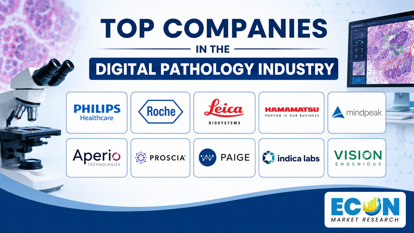
The following 10 companies are presented according to product capabilities, regulatory achievements, scanner and software expertise, geographic reach, clinical deployments and relevance to digital pathology. The order is not a strict ranking.
1. Royal Philips
Company Overview: Philips employs approximately 64,300 people and provides products and services in more than 100 countries. Headquarters: Amsterdam, Netherlands. Core Digital Pathology Expertise: The company develops complete diagnostic environments incorporating scanners, displays, image management, storage, interoperability and cloud deployment. Major Products and Services: Philips IntelliSite Pathology Solution includes second-generation pathology scanners, an image-management viewer, application server, storage software and a validated 27-inch display for applicable United States configurations. The system supports primary interpretation of formalin-fixed, paraffin-embedded tissue and conforms to European IVDR requirements. In 2026, Philips expanded IntelliSite through its HealthSuite cloud environment, strengthening multi-site access and scalable storage. Its open-platform approach also supports third-party AI integration, including expanded collaboration with Ibex announced in 2025.
2. Roche
Company Overview: Roche was founded in Basel in 1896 and provides medicines and diagnostics across more than 150 countries. Headquarters: Basel, Switzerland. Core Digital Pathology Expertise: Roche integrates tissue staining, whole-slide scanning, image management and computational analysis within one pathology-laboratory portfolio. Major Products and Services: Roche Digital Pathology Dx combines the VENTANA DP 200 and DP 600 scanners, uPath enterprise software, navify Digital Pathology and a validated display. The DP 600 received an expanded FDA clearance in January 2025 after the original system clearance in June 2024. Roche also develops image-analysis algorithms for biomarkers including HER2 and received FDA Breakthrough Device Designation in 2025 for an AI-driven TROP2 companion diagnostic. The open environment allows third-party analytical tools to connect with navify workflows using published scanner file specifications.
3. Leica Biosystems
Company Overview: Leica Biosystems operates as a Danaher company and maintains its international headquarters in Nussloch, Germany. Headquarters: Nussloch, Germany. Core Digital Pathology Expertise: The company combines slide preparation, staining, whole-slide imaging, image management and quantitative analysis across clinical and research workflows. Major Products and Services: Leica expanded its diagnostic portfolio in December 2025 with the Aperio GT 180 DX, Aperio CS5 DX and Aperio iQC DX software. The CS5 supports 1×3-inch and 2×3-inch slides, optional Z-stacking and 20× or 40× magnification. Leica also introduced the Aperio FL system in April 2025 for fluorescence imaging and launched Aperio HALO AP software later that year. Its scanner ecosystem supports eSlide Manager, WebViewer DX and integrations with AI products from specialist developers.
4. Hamamatsu Photonics
Company Overview: Hamamatsu Photonics employs over 6,600 people, serves more than 100 countries and operates subsidiaries in 16 countries and regions. Headquarters: Hamamatsu, Japan. Core Digital Pathology Expertise: The company applies its optical-sensor, camera and photonics capabilities to high-resolution whole-slide imaging. Major Products and Services: Hamamatsu’s NanoZoomer portfolio includes the S20MD, S360MD and S540MD clinical systems alongside research scanners. NZConnectMD Scan, introduced in April 2025, enables centralized, barcode-driven control of multiple scanners. India approved the S360MD and S20MD in July 2025, while several NanoZoomer systems hold Canadian medical-device licenses. Hamamatsu scanners integrate with third-party diagnostic viewers and AI platforms, including FDA-cleared combinations involving Paige FullFocus and Leica HALO AP Dx.
5. Sectra
Company Overview: Sectra was founded in 1978, employs approximately 1,100 people and maintains direct sales in 18 countries with partners serving additional markets. Headquarters: Linköping, Sweden. Core Digital Pathology Expertise: Sectra specializes in enterprise image management, integrated diagnostics, cloud infrastructure and cybersecurity rather than manufacturing slide scanners. Major Products and Services: The Sectra Digital Pathology Module combines worklists, reporting, image review and AI integration in the same enterprise platform used for radiology and other imaging specialties. The company marked 10 years of digital pathology activity before its Canadian go-live in January 2026. Its deployments include 5 NHS sites in Devon and Cornwall, 3 French university hospitals and Region Västra Götaland in Sweden. Sectra also distributes pathology AI through its Amplifier marketplace, including an agreement with Paige announced in April 2025.
6. Proscia
Company Overview: Proscia was founded in 2014 by a team connected with Johns Hopkins University, Moffitt Cancer Center and the University of Pittsburgh. Headquarters: Philadelphia, Pennsylvania, United States. Core Digital Pathology Expertise: The company develops cloud-native software for clinical diagnostics, pharmaceutical research, image management and AI deployment. Major Products and Services: Concentriq unifies whole-slide images, metadata, clinical history and study information within one platform. Its fifth generation, introduced in 2026, added an enterprise AI layer designed to bring models and experts into a shared operating environment. In September 2025, Proscia integrated Concentriq with AWS HealthImaging for standards-based DICOM storage at petabyte scale. Healthcare and laboratory customers use the platform for remote collaboration, case management, research and computational pathology, while partnerships with scanner companies provide integrated hardware-and-software deployment options.
7. PathAI
Company Overview: PathAI was founded in 2016 and maintains a network of more than 450 board-certified pathologists who have contributed 32.5 million annotations. Its technologies are used by 90% of the top 15 biopharmaceutical companies. Headquarters: Boston, Massachusetts, United States. Core Digital Pathology Expertise: PathAI combines image management, AI product development, clinical-trial pathology and computational biomarker analysis. Major Products and Services: AISight Dx received FDA clearance for primary diagnosis in June 2025 and supports scanner-agnostic workflows involving Hamamatsu, Leica, Roche and Philips systems. The platform offers case management, viewing, collaboration, DICOM support and AI integration. PathAI launched more than 20 AI products in 2023 and introduced its Precision Pathology Network in 2025. Applications address tumor cellularity, MASH, inflammatory bowel disease, tissue quality and tumor-microenvironment analysis.
8. Paige
Company Overview: Paige was founded in 2017 and developed its technology through collaboration with specialist cancer pathology institutions. Headquarters: New York City, United States. Core Digital Pathology Expertise: The company develops FDA-cleared viewing software, cancer-detection applications and large foundation models for computational pathology. Major Products and Services: FullFocus received FDA clearance for use with Leica Aperio GT 450 DX and Hamamatsu NanoZoomer S360MD scanners, supporting SVS, DICOM and NDPI file formats. Paige Prostate Detect became the first FDA-authorized AI application for cancer detection in pathology, while Paige Lymph Node received Breakthrough Device Designation for detecting breast-cancer metastases. Its Virchow model family includes a second-generation system trained with approximately 1 million slides. Paige also integrates third-party biomarker algorithms and distributes applications through enterprise partners such as Sectra, Lumea and OptraSCAN.
9. Ibex Medical Analytics
Company Overview: Ibex was founded in 2016 and operates from Tel Aviv and Boston as it expands clinical and biopharmaceutical activities. Headquarters: Tel Aviv, Israel. Core Digital Pathology Expertise: Ibex develops clinical-grade AI for detecting and grading cancers and quantifying biomarkers in routine pathology workflows. Major Products and Services: Its solutions address prostate, breast and gastric pathology and integrate with scanners, laboratory systems and image-management platforms from multiple suppliers. A CorePlus deployment had processed over 10,000 clinical cases by 2023, while a 2026 expansion introduced AI support for transurethral resection prostate specimens. Ibex obtained CE-IVDR certification in July 2026 for breast ER, PR and Ki-67 applications. Partnerships with Philips, Roche, Fujifilm, AWS and diagnostic laboratories support deployment across North America, Europe, Israel and Asia.
10. 3DHISTECH
Company Overview: 3DHISTECH was founded in 1996 and has sold more than 3,500 digital pathology systems. Headquarters: Budapest, Hungary. Core Digital Pathology Expertise: The company designs and manufactures whole-slide scanners, tissue-microarray systems, micro-CT instruments and software for clinical, research, toxicology and pharmaceutical applications. Major Products and Services: Its Pannoramic portfolio includes compact scanners and high-throughput systems such as the Pannoramic 480 and Pannoramic 1000. The Pannoramic 480 DX supports high-volume diagnostic workflows, while 10× objectives on selected scanners can reduce scanning time by half. The Pannoramic-X micro-CT platform images complete paraffin blocks and specimens measuring up to 30 centimeters without conventional sectioning. Software products include CaseManager DX, SlideManager, WebViewer and QuantCenter for case management and image analysis.
Regional Outlook
North America
North America combines established digital pathology regulation with large academic medical centers, reference laboratories and cancer networks. The United States authorized its first primary-diagnosis whole-slide imaging system in 2017 and continues to regulate scanners, viewers and computational tools through device-specific submissions. FDA clearances obtained during 2024 and 2025 covered Roche’s scanner environment, Paige’s multi-scanner viewer and PathAI’s AISight Dx platform. The region also contains the headquarters of at least 5 companies profiled in this article, supporting software development, clinical validation and regulatory expertise. United States adoption is strongest where laboratories can connect digital images with existing laboratory information systems, while reimbursement and operational governance continue to influence expansion beyond major centers.
Canada is moving from pilot projects toward routine deployment through provincial and hospital-level initiatives. Guelph General Hospital became the first Canadian hospital to activate Sectra’s Digital Pathology Module in January 2026 after beginning validation in September 2025; the hospital processes approximately 10,000 pathology examinations each year. Hamamatsu also holds Canadian medical-device licenses for 3 NanoZoomer scanner configurations. Mexico’s implementation remains more dependent on private laboratory networks, specialist hospitals and imported platforms than the regulated United States ecosystem. Across the 3 North American countries, the strongest opportunities involve remote subspecialty reporting, AI-supported cancer diagnosis and regional workload balancing. Key challenges include image-storage costs, cybersecurity, scanner maintenance and validating performance across diverse populations and laboratory procedures.
Europe
Europe has an extensive digital pathology manufacturing and clinical base, including Philips in the Netherlands, Roche in Switzerland, Leica Biosystems in Germany, Sectra in Sweden and 3DHISTECH in Hungary. The European Union comprises 27 member states operating under the In Vitro Diagnostic Medical Devices Regulation and the Artificial Intelligence Act. The AI Act entered into force in 2024, while the European Commission published guidance in June 2025 explaining its interaction with medical-device and in-vitro diagnostic requirements. Digital pathology companies must therefore coordinate quality management, clinical evidence, cybersecurity, post-market monitoring and high-risk AI obligations. This framework supports consistent safety expectations but can increase certification work for companies maintaining multiple scanners, viewers and algorithms.
The United Kingdom is an active implementation market despite operating outside the European Union framework. NHS England issued digital histopathology guidance in February 2025 for 3 national cancer-screening programs covering breast, bowel and cervical services. Sectra deployments connect 5 NHS sites in Devon and Cornwall, while its customer portfolio references digital pathology activity across 17 trusts in the West Midlands. Three French university hospitals signed digital pathology contracts in 2025, and Sweden’s second-largest healthcare region expanded its use of Sectra’s platform the same year. Europe’s main opportunities involve regional reporting networks, cloud services and cross-disciplinary cancer diagnostics. Challenges include different national procurement systems, long-term image retention and the availability of pathologists trained to validate and use digital systems.
Asia-Pacific
Asia-Pacific combines optical-manufacturing expertise, large diagnostic networks and expanding digital-health infrastructure. Hamamatsu Photonics maintains its headquarters and primary production and research facilities in Japan, operates through subsidiaries in 16 countries and regions, and supplies customers in over 100 countries. Its S360MD and S20MD scanners received Indian approval in July 2025, providing additional regulated options for laboratories digitizing histopathology workflows. Leica introduced the Aperio GT 180 at an Asia-Pacific digital pathology summit in August 2025, while 3DHISTECH distributes scanners across regional clinical and research markets. Japan, China, India, South Korea, Singapore, Taiwan and Australia provide distinct combinations of healthcare demand, manufacturing capability and regulatory requirements.
India’s large diagnostic chains create opportunities for AI-assisted review, illustrated by the deployment of Ibex technology within Dr. Lal PathLabs. Australia supports digital pathology through academic laboratories, regional care networks and scanner-software research, while Singapore provides a base for Southeast Asian medical-technology operations. China and Taiwan contribute imaging components, electronics and manufacturing capacity, but vendors must address local registration and data-governance requirements. Across 8 major Asia-Pacific markets, adoption will depend on scanner affordability, local service coverage, multilingual interfaces and reliable cloud connectivity. Regional laboratories may prioritize compact scanners and remote consultations before implementing full enterprise systems, while national reference laboratories can justify high-throughput automation and AI-based quality control.
Middle East and Africa
The Middle East is building a stronger digital-health foundation through national transformation programs. Saudi Arabia’s Digital Health Center of Excellence was launched in the third quarter of 2020, while Vision 2030 programs seek to connect healthcare information systems and support advanced diagnostic and decision tools. By July 2026, the Ministry of Health’s innovation sandbox had supported 342 digital-health solutions and graduated 25 after testing and validation. These programs create a pathway for digital pathology platforms, AI cancer diagnostics and remote specialist consultation. The 6 Gulf Cooperation Council countries can use cloud-based pathology to connect tertiary cancer centers with regional hospitals, but deployments require Arabic-language workflows, local data hosting and trained laboratory personnel.
Africa’s opportunity is closely linked to cancer-access and workforce limitations. WHO projects a regional shortage of 6.1 million health workers by 2030, and diagnostic competency assessments show an average accuracy of approximately 60% across evaluated healthcare workers. Digital pathology cannot replace tissue processing, staining or trained pathologists, but it can connect scarce experts with hospitals that lack subspecialty coverage. South Africa, Egypt, Kenya, Nigeria and Morocco have stronger laboratory and academic infrastructure than many neighboring markets, creating potential regional reporting hubs. Sustainable deployment must address electricity, scanner servicing, internet bandwidth and the cost of storing whole-slide images. Small laboratories may begin with telepathology and consultation workflows before adopting AI or fully digital primary diagnosis.
Future Opportunities in the Digital Pathology Industry
Future opportunities for the Top Companies in the Digital Pathology Industry will center on multimodal AI, workflow automation and open enterprise platforms. Foundation models trained on 500,000 to 1 million slides demonstrate the technical scale now available for research, but clinical applications will need focused validation for specific diseases, stains and patient populations. AI can assist with tumor detection, grading, cell counting, biomarker scoring and tissue selection for molecular testing. Future platforms may combine histology with radiology, genomics and laboratory results to create a more complete diagnostic view. Human oversight will remain necessary because tissue artifacts, staining variation and unusual tumor patterns can produce errors that require specialist interpretation.
Cloud and edge infrastructure will support larger regional deployments through 2030. Standards such as DICOM can reduce dependence on proprietary formats, while petabyte-scale storage services can manage growing archives across networks containing 10 or more laboratories. Automated quality control can identify blurred tissue, missing sections and scanning artifacts before cases reach pathologists. Barcodes, robotic slide handling and centralized scanner control can reduce repetitive work and improve traceability. Smaller digital pathology developers can reach international customers through partnerships with scanner manufacturers and enterprise imaging providers rather than building independent distribution in 100 countries. Regulatory success will depend on predefined change controls, transparent algorithm updates and continuous monitoring after installation.
Sustainability and workforce development represent additional opportunities. Remote reporting can reduce physical transportation of slides between hospitals, while shared scanners and cloud infrastructure can improve equipment utilization across multi-site networks. Laboratories will still retain physical blocks and slides for defined periods; one NHS region maintains archives extending beyond 30 years, demonstrating that digital storage complements rather than immediately replaces physical archives. Training programs must prepare pathologists, technicians and IT teams to validate scanners, manage images and evaluate AI outputs. Companies that combine technology with implementation services, competency assessment and long-term support will be better positioned than suppliers offering hardware or algorithms without workflow transformation.
Conclusion
The digital pathology industry has moved from research-oriented slide scanning toward regulated primary diagnosis, enterprise image management and AI-assisted cancer assessment. The first United States whole-slide imaging authorization in 2017 created a foundation for newer scanners, viewers and cloud platforms cleared during 2024 and 2025. The 5 defining trends—cloud interoperability, foundation models, integrated diagnostics, high-throughput automation and lifecycle governance—are changing how pathology laboratories manage cases and collaborate. The 10 selected companies contribute different capabilities across scanning, image storage, workflow software, artificial intelligence, biomarker analysis and multi-site deployment.
Regional conditions remain uneven. North America has a developed FDA pathway, Europe combines 27 European Union markets with emerging high-risk AI obligations, Asia-Pacific provides manufacturing and diagnostic scale, and the Middle East and Africa are building digital infrastructure around workforce and access needs. The strongest opportunities involve open systems, DICOM adoption, AI quality control, remote subspecialty reporting and multimodal cancer diagnostics. The Top Companies in the Digital Pathology Industry will ultimately be differentiated by their ability to maintain image quality, regulatory compliance, interoperability, cybersecurity and clinical trust across thousands of scanners, users and diagnostic cases.